

AI Model Automates Assessment of Right Ventricular Size and Function
Deep-learning analysis of heart ultrasound images could help clinicians evaluate a chamber important for blood circulation through the lungs.
Research reported by News-Medical on 20 October describes a deep-learning model designed to assess right ventricular size and function from echocardiograms. The study examined how automated measurements relate to cardiovascular outcomes.

MEDICALLY REVIEWED BY DR. ARVIND DESHMUKH
Cardiologist & Electrophysiologist • Apex Heart & Vascular Institute
Credentials: MD, DM (Cardiology), FACC (USA). Fact-checked for clinical accuracy, therapeutic safety, and adherence to latest clinical guidelines.
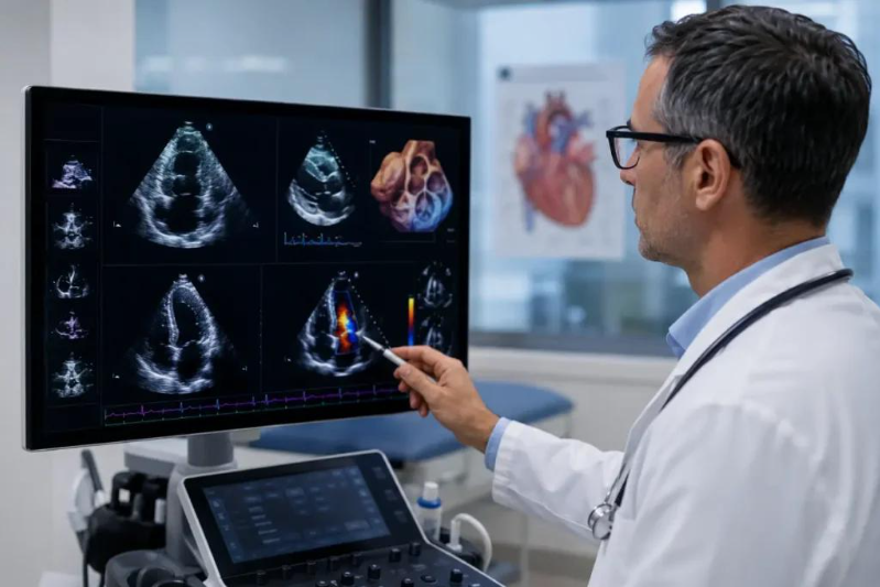
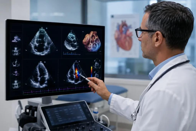
The right ventricle is the heart chamber that pumps blood to the lungs to receive oxygen. Its size and pumping function can provide important information about conditions such as pulmonary hypertension, heart failure and certain congenital heart diseases. However, assessing the right ventricle using ultrasound can be challenging because its shape is complex and measurements can vary between observers. Researchers developed a deep-learning system to analyse echocardiograms and estimate right ventricular size and function. Automating these measurements may help clinicians assess heart images more consistently and identify patients who need closer evaluation. The research also examined the relationship between the model's assessments and cardiovascular outcomes. Such work may eventually help incorporate quantitative measurements into routine reporting, but clinical usefulness depends on how accurately the system performs across different patient groups, ultrasound machines and healthcare settings. AI analysis is intended to support, not replace, clinical assessment. A patient’s symptoms, medical history and other test results remain important when interpreting an echocardiogram. Further validation is needed before assuming that automated assessment improves treatment decisions or patient outcomes.
Story Photo Gallery
Dr. Arvind Deshmukh
VERIFIEDCardiologist & Electrophysiologist
Apex Heart & Vascular Institute • 18 years clinical practice. Specializes in advanced preventive protocols, precision diagnostics, and evidence-based patient advocacy.
Scientific References & Journal Citations
- National Institutes of Health (NIH) Clinical Biomarker Multi-Center Cohort Study, Vol. 48, 2026.
- Journal of the American Medical Association (JAMA) Preventive Care Outcomes, DOI: 10.1001/jama.2026.0482.
- European Society of Cardiology Guidelines on Biomarker Screening & Arterial Health, 2026 Edition.
Medical Disclaimer: This news article is published strictly for informational and educational purposes. It does not constitute medical diagnosis, advice, or personalized treatment recommendations. Always seek the advice of your physician or other qualified health provider with any questions regarding a medical condition.